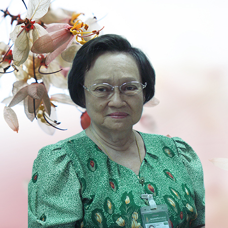
- 1962 Doctor of Medicine (M.D.) Faculty of Medicine Siriraj Hospital, College of Medicine
- 1963 Internship training program, Faculty of Medicine Siriraj Hospital, with Medical License and Higher Graduate Diploma in Medicine
- 1967 MSc. Virology Department, Royal Children’s Hospital, Melbourne University, Australia
- 1968 - 1970 Lecturer, Department of Microbiology, in Virology Faculty of Medicine Siriraj Hospital, College of Medicine, Bangkok, Thailand
- 1970 - 1979 Assist Prof, Department of Anatomy, Faculty of Medicine Siriraj Hospital, College of Medicine, Mahidol University (1971) Bangkok, Thailand
- 1979 - 1998 Assist Prof, Department of Anatomy, Faculty of Science, Medicine Prince of Songkhla University Hat Yai, Songkhla, Thailand
- 1998 - 1999 Lecturer, Professional level, Department of Anatomy Faculty of Science, Medicine Prince of Songkla University Hat Yai, Songkhla, Thailand
- 1999 - 2009 Lecturer, Professional level, Medical Education Section, Faculty of Medicine, Prince of Songkla University Hat Yai, Songkhla, Thailand
- 1978 – 1979 Composing text on anatomy "Atlas of Human Histology" (18 Chapters,with 287 color pictures,twice publication, 4000 volumes)
- 2001 - 2003 Design of Photography Study of Human Body/Anatomy (Gross, Histology, Embryology Neuroanatomy + Topographic anatomy) in Dunuclear Transparency, Permanently attached to the three walls of the museum. (298 color illustrations with captions)
- 1999 - 2001 - present Design of CAI teaching materials on anatomy covering all divisions being in line with Medical curriculum of Block System:
- 1. Anatomy of the Heart (Gross Anatomy) (1999; 3 sections, with 52 colors illustrations)
- 2. Placenta and Fetal Membranes (Embryology) (2001; 3 sections, with 52 color illustrations)
- 3. Development of Integumentary System (Embryology) (2002; 4 sections, with 22 color illustrations)
- 4. Integumentary System ( Histology )(2003; 5 sections, with 88 color illustrations)
- 5. Respiratory System (2004; 2 Volumes, with 121 color illustrations)
- - Volume 1 Conducting Portion (Histology) (9 sections, with 62 color illustrations)
- - Volume 2 Bone (Histology) (9 sections, with 59 color illustrations)
- 6. Skeletal System.(2005; with 122 color illustrations, 3 volumes)
- - Volume 1 Cartilage (Histology) (8 sections, with 31 color illustrations)
- - Volume 2 Bone (Histology) (8 sections, with 42 color illustrations)
- - Volume 3 Osteogenesis and Growth (Histology) (8 sections, with 49 color illustrations)
- 7. Hematopoietic System(2006; with 193 color illustrations, 2 volumes)
- - Volume 1 Blood and Formed Elements (Histology)(6 sections, with 55 color illustrations)
- - Volume 2 Bone Marrow and Hematopoiesis (Histology)(7 sections, with 138 color illustrations)
- 8. Cardiovascular System ( 2007; with 159 color illustrations, 2 volumes)
- - Volume 1 Heart (Histology) (7 sections, with 77 color illustrations)
- - Volume 2 Blood Vessels : Arteries & Veins including Lymph Vessels (Histology) (4 sections, with 82 color
illustrations)
- 9. CAI : NEUROANATOMY-Test & Atlas. 10 Volumes, 44 Chapters
- - Volume 1 Organization of Nervous System (5 Chapters, 22 sections, with 200 color illustration)
- - Chapter 1 Central & Peripheral Nervous System : CNS & PNS (4 sections, with 47 color illustrations)
- - Chapter 2 Autonomic Nervous System (ANS) & Visceral Afferents (5 sections, with 20 color illustrations)
- - Chapter 3-4-5 Development & Histogenesis of NERVOUS SYSTEM :
- A. Development of Spinal Cord (4 sections, with 26 color illustrations)
- B. Development of Brain (5 sections, with 87 color illustrations)
- C. Development of PNS & ANS (4 sections, with 20 color illustrations)
- To produce CAI NEUROANATOMY (Text&Atlas) 10 volumes, 44 chapters.
- To produce CAI HISTOLOGY (Atlas) 18 Chapters, 292 color illustrations, with captions.
- To produce CAI EMBRYOLOGY : Development of CARDIOVASCULAR SYSTEM (Block 3).
Primarily, the production of CAI teaching materials means to increase basic knowledge of Anatomy in all levels, which has been reduced in the new Medical Science program : the BLOCK SYSTEM, as well as to enhance complete knowledge, equivalent to other Medical Schools (Siriraj, Khonkhen, Chiangmai etc) whose Medical programs still rely on conventional Medical Science curriculum focusing on lectures, practice and cadaveric dissections throughout the academic year.

